4周搞定2000+托福核心词
microcosm = micro(微,小) + cosm(宇宙) = 微观世界
microbiologist = micro(微,小) + bi(生物) + ology(学科) + ist(人) = 微生物学家
scope = scope(视线) =
stereoscopeic = stereo(立体的) + scope(视线) + ic(…的) = 立体的;体视镜的
看小东西→显微镜
microscope
常考释义
This system, organizing all life into these two kingdoms, worked very well for quite a while, even into the age of the microscope.
这个分类系统把所有的生命体都划分进了这两个界,它一直起着很好的分类作用,即使是在发明了显微镜的时代。
来源于:听力OFFICIAL50 L2n. 显微镜
Viewing a section of the spinal cord under the microscope, Cho was amazed to see that the spinal cord was completely dark.
在显微镜下看一个脊髓节,赵很惊讶地看到,脊髓完全黑了。
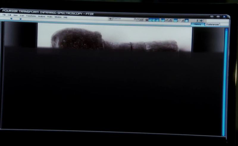
I put our bullet under electron microscope magnification.
我把弹头放在电子显微镜下进行放大。

来自《犯罪现场调查 第13季第9集》
图片源自网络
All you do is to train the microscope on tiny flecks of paint and analyze them.
你所做的就是;练习使用显微镜对准油画上的小小斑点,进行分析。
来源于:听力OFFICIAL5 L5Well, we put an infrared microscope - a spectroscope - on tiny tiny bits of paint.
嗯,我们使用红外线显微镜,一个分光镜,来观察油画上很小很小的部分。
来源于:听力OFFICIAL5 L5I was so impressed with the way you handled the microscope and the samples of onion cells and, well, how careful you observed and diagramed and interpreted each stage of cell division, and I don’t think you could have done that if you hadn’t understood the chapter.
你操作显微镜以及洋葱细胞样本的方式让我很印象深刻,你很认真的观察、画图并且解释了细胞分裂的每个过程。我想如果你没有读并理解这个章节的内容的话,也不会做的那么好。
来源于:听力OFFICIAL15 L4The size of a cell often determines the type of microscope a biologist uses to study it.
细胞的大小经常决定了生物学家选用哪种显微镜来研究它。
来源于:阅读OFFICIAL38 P1Thus, the light microscope remains a useful tool, especially for studying living cells.
因此,光学显微镜仍是一个有用的工具,尤其是研究活的细胞的时候。
来源于:阅读OFFICIAL38 P1For a biologist studying a living process, such as the whirling movement of a bacterium, a light microscope equipped with a video camera might be better than either a scanning electron microscope or a transmission electron microscope.
如果一个生物学家正在研究一个活体过程,比如一个细菌的旋动,装备有摄像机的光学显微镜就比扫描电子显微镜或透射电子显微镜好用了。
来源于:阅读OFFICIAL38 P1Electron microscopes have truly revolutionized the study of cells and cell organelles.
电子显微镜的确彻底变革了对细胞和细胞器的研究。
来源于:阅读OFFICIAL38 P1However, instead of lenses made of glass, the transmission electron microscope uses electromagnets as lenses, as do all electron microscopes.
但是,与光学显微镜不同的是,它不是用玻璃作为镜片,而是使用电磁体作为镜片,正如所有的电子显微镜一样。
来源于:阅读OFFICIAL38 P1Specimens are cut into extremely thin sections, and the transmission electron microscope aims an electron beam through a section, just as a light microscope aims a beam of light through a specimen.
样本被切割成极其薄的切片,透射电子显微镜将电子束穿透切片,正如光学显微镜将光束透过样本一样。
来源于:阅读OFFICIAL38 P1The transmission electron microscope, on the other hands, is used to study the details of internal cell structure.
另一方面,透射电子显微镜被用来研究细胞的内部结构。
来源于:阅读OFFICIAL38 P1The scanning electron microscope produces images that look three-dimensional.
扫描电子显微镜的成像看起来是三维立体的。
来源于:阅读OFFICIAL38 P1Biologists use the scanning electron microscope to study the detailed architecture of cell surfaces.
生物学家使用扫描电子显微镜研究细胞表面的具体构造。
来源于:阅读OFFICIAL38 P1Only under special conditions can electron microscopes detect individual atoms.
只有在特殊情况下电子显微镜才能发现原子。
来源于:阅读OFFICIAL38 P1In fact, the most powerful modern electron microscopes can distinguish objects as small as 0.2 nanometers, a thousandfold improvement over the light microscope.
实际上,现代最先进的电子显微镜能够识别直径为0.2纳米的物体,比光学显微镜进步了成千倍。
来源于:阅读OFFICIAL38 P1Instead of light, the electron microscope uses a beam of electrons and has a much higher resolving power than the light microscope.
不再利用光线,电子显微镜利用的是电子束而且比光学显微镜具有更高的分辨率。
来源于:阅读OFFICIAL38 P1Our knowledge of cell structure took a giant leap forward as biologists began using the electron microscope in the 1950s.
直到二十世纪50年代,由于电子显微镜的使用,我们对于细胞结构的了解实现了巨大飞跃。
来源于:阅读OFFICIAL38 P1From the year 1665, whenEnglish microscopist Robert Hooke discovered cells, until the middle of thetwentieth century, biologists had only light microscopes for viewing cells.
从1665年英国显微镜学家Robert Hooke发现细胞到二十世纪中期,生物学家只能通过光学显微镜来观察细胞。
来源于:阅读OFFICIAL38 P1The light microscope cannot resolve detail finer than 0.2 micrometers, about the size of the smallest bacterium; consequently, no matter how many times its image of such a bacterium is magnified, the light microscope cannot show the details of the cell's internal structure.
光学显微镜不能够分辨小于0.2微米的细节,这相当于最小的细菌的大小;因此,不论这个细菌被放大了多少倍,光学显微镜都不能展现出细胞内部结构的细节。
来源于:阅读OFFICIAL38 P1Light microscopes can magnify objects up to1,000 times without causing blurriness.
光学显微镜能将物体放大1000倍而不会模糊不清。
来源于:阅读OFFICIAL38 P1Glass lenses in the microscope bend the lightto magnify the image of the specimen and project the image into the viewer'seye or onto photographic film.
显微镜中的玻璃镜片通过弯曲光线将样本放大,并将样本的影像投进观察者的眼中或者感光胶片上。
来源于:阅读OFFICIAL38 P1The first microscopes were light microscopes, which work by passingvisible light through a specimen.
第一代显微镜是光学显微镜,其工作原理是将可视光透过样本。
来源于:阅读OFFICIAL38 P1Before microscopes werefirst used in the seventeenth century, no one knew that living organisms werecomposed of cells.
在17世纪显微镜第一次投入使用之前,人们都不知道生物体是由细胞构成的。
来源于:阅读OFFICIAL38 P1And as microscopes improved, we discovered some microorganisms that were incredibly small.
随着显微镜的逐渐改进,我们发现一些微生物是非常之小的。
来源于:听力OFFICIAL50 L3But using the microscope we discovered that a fungus contains these microscopic thread-like cells that run all over the place.
但是使用显微镜,我们发现真菌身上有这些微小的线状细胞,它们到处都是。
来源于:听力OFFICIAL50 L3A hundred years or so after the invention of the microscope, Carolus Linnaeus devised a simple and practical system for classifying living things, according to the ranks of categorization still in use today——class, order, family and so on.
大约在显微镜发明的一百年后,卡罗勒斯•林奈发明出了一个给生物分类的简单而实用的系统,这个系统是根据等级编目法的——涉及的等级有纲,目,科等等,这些等级现在还在使用。
来源于:听力OFFICIAL50 L3With the invention of the microscope, in the late 1500s, we discovered the first microorganisms.
随着十六世纪末期显微镜的发明,我们发现了第一批微生物。
来源于:听力OFFICIAL50 L3This system, organizing all life into these two kingdoms, worked very well for quite a while, even into the age of the microscope.
这个分类系统把所有的生命体都划分进了这两个界,它一直起着很好的分类作用,即使是在发明了显微镜的时代。
来源于:听力OFFICIAL50 L3n. under the microscope:被仔细检查
The media put their every decision under the microscope.
媒体非常仔细地研究了他们的每一项决定。
He ignored the question. I was the one under the microscope.
他没理这个问题,我成了仔细盘问的对象。
electron microscope
电子显微镜
以上图片仅供学习交流使用,版权归原作者所有,如有侵权,请与我方联系